
Technical Review: FAP (Prolyl Endopeptidase) ELISA – Applications, Methodology & Research Utility
FAP ELISA, fibroblast activation protein alpha assay, prolyl endopeptidase FAP, FAP biomarker, ELISA protein separation, FAP immunoassay kit, stromal fibroblast marker FAP, tumor microenvironment FAP, FAP activity assay, FAP diagnostic biomarker.
Introduction and Biological Context
The enzyme known as fibroblast activation protein alpha (FAP-α) is a type II transmembrane serine protease of the S9B prolyl oligopeptidase family, with dual dipeptidyl peptidase and endopeptidase activities. aacrjournals.org+3PubMed+3PubMed+3 FAP is encoded by the gene FAP (Gene ID: 2191) in Homo sapiens. CNIB
Functionally, FAP has been implicated in tissue remodelling, extracellular matrix (ECM) degradation, wound healing, fibrosis, and most notably, tumour-associated stroma in many epithelial cancers. CNIB+1 Because of its restricted expression in adult normal tissues but high expression in activated fibroblasts (cancer-associated fibroblasts, CAFs) and areas of ECM turnover, FAP has emerged as a promising biomarker and target for diagnostics and therapeutics. PubMed+1
In the immunoassay context, an ELISA (enzyme-linked immunosorbent assay) for FAP enables quantitative measurement of its antigen (protein) levels — either in soluble form (shed or secreted FAP) or in sample extracts (serum, plasma, conditioned medium). Kits are commercially available for “FAP ELISA” as well as for the related enzyme Prolyl Endopeptidase (PREP) (alias “prolyl oligopeptidase”). biocompare.com+1
Thus, a robust FAP ELISA workflow allows research labs in oncology, fibrosis, immunology and biomarker development to quantify FAP levels with high sensitivity and specificity.
![AffiELISA® Mouse Prolyl endopeptidase FAP ELISA [ Fap]](https://affigen.com/cdn/shop/files/5BAFG-E4345_5D_20AffiELISA_C2_AE_20Cattle_20IFNg_20Kit_20High-Resolution_20Interferon_20Gamma_20ELISA_20Detection_ed6b212c-d0a0-485c-a506-bf8c31769b64_535x.png?v=1712852291)
Enzyme Biochemistry & Substrate Specificity
Catalytic Mechanism
FAP comprises an extracellular catalytic domain with a classical serine-protease catalytic triad (Ser^624, Asp^702, His^734 in human sequence) and an eight-bladed β-propeller domain that restricts substrate entry, conferring its specificity. PubMed+1
FAP can cleave substrates at the post-proline bond (-Pro-Xaa) (endopeptidase) and also exhibits dipeptidyl peptidase activity (cleaving Xaa-Pro motifs). This dual specificity sets it apart from other prolyl-cleaving proteases such as DPPIV/CD26. PubMed+1
For example, FAP has been shown to cleave the metabolic hormone FGF21 in vitro and to degrade denatured collagen I/III in stromal matrix contexts. CNIB
Expression and Tissue Distribution
Under physiological (normal adult) conditions, FAP expression is very low or absent in most organs. In contrast, FAP is highly expressed in activated stromal fibroblasts during wound healing, fibrotic diseases, and the tumour microenvironment (TME). PubMed+1 The restricted basal expression and elevated pathological expression make FAP especially useful as a biomarker.
Functional Implications
Because of its ability to degrade ECM components and modify the stromal environment, FAP contributes to matrix remodelling, fibroblast migration, tumour invasion, immunosuppression in the TME and fibrosis progression. PMC+1
From a biomarker standpoint, measuring the soluble or shed FAP antigen in plasma or serum can provide insights into the fibroblast-activation status, stromal remodelling or disease progression.
ELISA Assay Principles for FAP
Sandwich ELISA Format
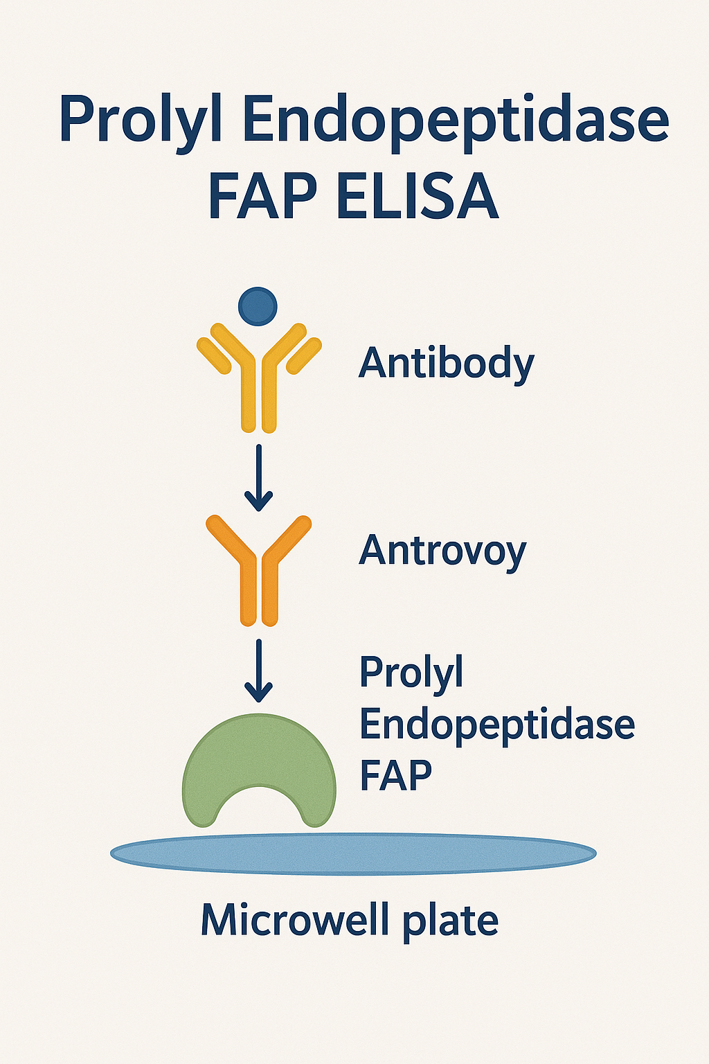
Most FAP ELISA kits are of the sandwich format: they use a capture antibody coated on the microplate, sample (containing FAP) is added, then a biotinylated or enzyme-conjugated detection antibody binds the captured FAP, followed by a substrate reaction (e.g., TMB for HRP) and optical read-out at ~450 nm. Examples: The Human FAP ELISA Kit (Abcam ab256404) uses a single-wash 90-min protocol, sensitivity ~14.96 pg/mL. abcam.com
Sample Types
Typical sample matrices: human serum, plasma (EDTA, heparin, citrate), cell culture supernatant, tissue homogenates. The protocol may require dilution of the sample to bring the FAP concentration into the measurable range. abcam.com+1
Standard Curve and Quantitation
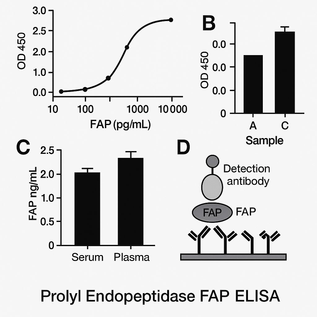
The kit includes a standard recombinant FAP calibrator. A standard curve is generated (e.g., serial dilutions from ~0.01 ng/mL to several ng/mL). Sample optical densities are interpolated against the standard curve to derive FAP concentrations.
Linearity, spike recovery, intra- and inter-assay coefficients of variation (CV) are key performance metrics; the Abcam kit reports CVs ~5-7 % within assay, ~10 % between assays. abcam.com
Sensitivity and Specificity
High-sensitivity kits claim lower limits of detection (LLOD) in the pg/mL range (e.g., 9.01 pg/mL for R&D Systems Human FAP QuicKit ELISA). www.rndsystems.com Specificity is achieved via validated monoclonal antibodies and minimal cross‐reactivity with related proteases (e.g., DPPIV, PREP).
Assay Time and Throughput
Assay times typically range from 1 hour (rapid kits) to 4 hours (standard kits). Given 96-well plates and automation potential, throughput is suitable for research labs and biomarker screening.
Methodology: Protocol Workflow & Best Practices
Pre-Analytical Considerations
-
Use fresh or properly stored (-80 °C) plasma/serum samples; avoid repeated freeze-thaw cycles.
-
Clarify samples by centrifugation to remove particulates.
-
Dilute samples in kit diluent as prescribed (commonly 1:5 to 1:50) to ensure readings fall within the linear range of the standard curve.
Plate Setup
-
Allow plate, reagents and samples to equilibrate at room temperature (~20-25 °C).
-
Add standards, blanks and samples in replicates (usually duplicates or triplicates) to wells.
Incubation & Detection
-
Incubate capture antibody‐coated wells with sample for specified time (e.g., 60 min).
-
Wash thoroughly (3-5 washes) with wash buffer to remove unbound material.
-
Add detection antibody, incubate as specified (e.g., 30 min).
-
Add enzyme conjugate (if separate), followed by substrate (TMB/H₂O₂).
-
Stop the reaction with acid (e.g., 1 M H₂SO₄) and read at 450 nm (with 570 or 630 nm reference).
Data Analysis
-
Generate standard curve (OD vs concentration) using 4- or 5-parameter logistic (4PL/5PL) regression.
-
Interpolate sample OD values to derive FAP concentration.
-
Correct for sample dilution factor.
-
Report mean ± standard deviation of replicate wells.
Quality Control and Troubleshooting
-
Include internal quality controls (low, medium, high concentration).
-
Verify recovery of spiked samples (expected 90-110 %).
-
Check linearity of diluted samples (dilution linearity within ±10 %).
-
If high background: reduce incubation time, increase washing, check reagent contamination.
-
If low signal: verify plate reader settings, reagent expiry, sample integrity.
Research Applications and Use Cases
Cancer Biomarker Research
Given FAP’s overexpression in tumour stroma and its role in invasion and metastasis, FAP antigen quantification by ELISA assists in:
-
Profiling stromal activation in tumour biopsies and conditioned media.
-
Evaluating soluble FAP levels as a circulating biomarker of extent of stromal reaction and possible prognostic value (studies show high FAP correlates with poorer outcomes). Frontiers
-
Screening anti-FAP therapeutic strategies and monitoring target engagement.
Fibrosis and Wound-Healing Research
FAP is up-regulated in fibrotic tissue (e.g., liver, lung) and in wound granulation fibroblasts. The ELISA allows quantitation of FAP levels in tissue extracts or plasma in models of fibrosis or wound repair. PMC
Drug Discovery & Inhibitor Testing
FAP inhibitors (small molecules or antibodies) require quantification of FAP antigen or activity for pharmacodynamic read-outs. ELISA enables high-throughput screening of samples from treated cell lines, animal plasma, or human cohorts.
Stromal Microenvironment Studies
In stromal biology, the level of FAP correlates with fibroblast activation status, ECM remodelling, myofibroblast differentiation and CAF phenotype. Measuring FAP can complement gene expression (mRNA) and immunohistochemistry (IHC) data.
Comparative Enzyme Activity vs Antigen Quantitation
While ELISA measures antigen concentration of FAP, enzyme-activity assays (fluorometric or chromogenic) measure functionality (proteolytic activity). It is often insightful to correlate antigen levels (via ELISA) with enzymatic activity (via substrate assays) to assess whether elevated antigen corresponds to active protease. For example, a “Selective Homogeneous Assay for Circulating Endopeptidase FAP” was described in Sci Rep. Nature
Similarly, an assay for prolyl endopeptidase (PREP) in serum used a chromogenic substrate Z-Gly-Pro-p-nitroanilide. bio-protocol.org+1
Such dual-read strategies strengthen biomarker workflows.
Advantages and Limitations of FAP ELISA
Advantages
-
High sensitivity, often pg/mL detection limits (e.g., 9 pg/mL in marketed kits). www.rndsystems.com
-
High specificity with monoclonal antibodies validated against related proteases.
-
Quantitative and reproducible across plates and runs.
-
Versatility across sample types (serum, plasma, culture supernatant, tissue extracts).
-
Relatively rapid turnaround (1–4 hours) when compared to other biomarker assays.
Limitations / Considerations
-
Antigen vs activity disconnect: ELISA measures protein amount but not necessarily active enzymatic form; inactive or truncated protein may still be detected.
-
Soluble vs membrane-bound forms: FAP is membrane-bound but may shed or be secreted in soluble form; assay must be validated for the relevant isoform.
-
Pre-analytical variability: Sample matrix effects (hemolysis, lipemia, anticoagulants) may influence results — rigorous validation required.
-
Cross-reactivity and interference: Related proteases or endogenous inhibitors may interfere unless the assay is well validated.
-
Biological heterogeneity: Because FAP is a stromal marker, levels may vary widely depending on tissue type, disease state, sample collection site and time. Thus, establishing reference ranges for specific patient cohorts is necessary.
Protocol Optimization and Practical Tips
-
Dilution factor selection: Run preliminary dilutions (e.g., 1:5, 1:10, 1:50) to figure out approximate concentration so that OD lies in mid-range of standard curve.
-
Duplicate/triplicate wells: Helps reduce assay variability and allows calculation of CVs.
-
Avoid edge-effects: Do not load edge wells first/last; ensure plate sits flat and stable during incubation.
-
Washing stringency: Insufficient washing can increase background; automated plate washers help.
-
Calibration curve check: Each run should include full standard curve; if slope or intercept shifts significantly, results should be discarded or repeated.
-
Data correction: Use blank well OD to subtract non-specific binding.
-
Matrix compatibility: If sample matrix (e.g., plasma) differs from standard curve diluent, perform spike recovery and dilution linearity tests to confirm no matrix effect.
-
Storage and stability: Store kits per manufacturer instructions (typically 2-8 °C unopened); once opened, use components within recommended time and avoid freeze-thaw cycles.
-
Standardisation for publication: Report key metrics: sensitivity, LLOQ (lower limit of quantitation), intra- and inter-assay CVs, recovery %, linearity %, sample types, dilution factor. This enhances comparability across studies.
Data Interpretation & Biomarker Strategy
When interpreting results from a FAP ELISA, consider:
-
Absolute concentration: e.g., ng/mL of FAP in plasma; compare with normal/control cohort to determine fold-change.
-
Change over time: In longitudinal studies, monitoring FAP levels pre- and post-treatment provides dynamic insight.
-
Correlation with other markers: Combine FAP data with CAF markers (e.g., α-SMA, PDGFRβ), matrix remodelling markers (MMPs, collagen fragments) or tumour biomarkers for richer profiling.
-
Clinical cut-offs: For translational studies, determine cut-off values (e.g., ROC curves) to segregate high vs low FAP expression groups.
-
Biological context: Elevated FAP may reflect stromal activation rather than tumour cell expression; interpret in conjunction with tissue context and cell-type specificity.
-
Activity vs antigen: If enzyme activity assays are run in parallel, discrepancy between antigen amount and activity may signal presence of inactive isoforms or inhibitors.
Regulatory and QA Considerations
For translational or diagnostic use of FAP ELISA:
-
Validated SOPs and documentation of assay performance (sensitivity, specificity, precision) are essential.
-
Reference reagents and batch tracking support reproducibility.
-
If samples are human clinical specimens, ensure IRB/ethical approval and sample handling according to biobank standards.
-
Laboratories should follow GLP/CAP/CLIA guidelines as applicable (for US: Clinical Laboratory Improvement Amendments — CLIA standards).
-
Data reporting should include lot numbers, operator, date, assay version, plate map and raw OD values (for audit trail).
Future Directions and Emerging Trends
-
Multiplex biomarker panels: Integrating FAP ELISA with soluble CAF markers, ECM fragments, and immune checkpoint proteins for comprehensive microenvironment profiling.
-
Companion diagnostics: Using FAP levels to stratify patients for anti-FAP therapies (e.g., CAR T, antibody-drug conjugates) or stromal-targeting agents.
-
Point-of-care / microfluidic versions: Efforts to miniaturise FAP detection for rapid bedside assays.
-
Activity-based assays linkage: Combining antigen ELISA with proteolytic activity read-outs to refine biomarker specificity (see homogeneous FAP activity assay as above). Nature
-
Longitudinal cohorts and real-world data: Establishing normal reference ranges, population variability and disease-specific cutoff ranges for soluble FAP in plasma in fibrosis, cancer, cardiovascular remodeling.
-
Regulatory approval pathway: Transitioning FAP ELISA from research use only (RUO) toward IVD (in vitro diagnostic) classifiers, necessitating clinical validation studies.
Summary
In summary, the FAP (prolyl endopeptidase FAP) ELISA is a robust, high-sensitivity immunoassay that enables quantitative measurement of a key stromal biomarker implicated in tumour biology, fibrosis and tissue remodelling. Its advantages—high specificity, versatile sample types, and quantitative read-out—make it a valuable tool for research labs, biomarker development and potentially translational diagnostics.
However, careful attention to pre-analytical variables, assay validation parameters, and data interpretation is essential to derive meaningful conclusions. As the field evolves, FAP ELISA will increasingly form part of biomarker panels and stromal-targeting strategies in oncology, regenerative medicine and fibrosis research.
References for Further Reading
-
Gene summary of FAP: NCBI Gene ID 2191 CNIB+1
-
Hamson EJ et al. “Understanding fibroblast activation protein (FAP): substrates, activities…” Proteomics Clin Appl. 2014. PubMed
-
Kelly T et al. “Fibroblast activation protein-α: a key modulator…” Int Rev Cell Mol Biol. 2012. PubMed
-
Bainbridge TW et al. “Selective Homogeneous Assay for Circulating Endopeptidase FAP” Sci Rep. 2017. Nature
-
Bio-protocol: “Prolyl Endopeptidase Assay (96-well plate)” – protocol for related enzyme PREP. bio-protocol.org
-
Biocompare listing: FAP ELISA kits for research use. biocompare.com
-
Abcam product page: Human FAP ELISA Kit details. abcam.com